6-OHDA Lesion in Rat
Discover how Melior’s unique phenotypic screening platforms can uncover the untapped value of your candidate therapeutic
The neurotoxin 6-hydroxydopamine (6-OHDA) is widely used to induce depletion of dopaminergic neurons in animal models of Parkinson’s Disease (PD). Unilateral administration of 6-OHDA into the medial forebrain bundle can produce a 90-95% ipsilateral (lesioned-side) depletion of dopamine neurons in 80-90% of animals injected, leading to a PD – like motor dysfunction.
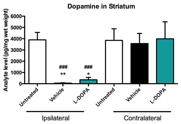
Striatal dopamine in the 6-OHDA model. Striatal dopamine was significantly reduced in vehicle and L-DOPA treated animals compared to unlesioned (untreated) animals in the ipsilateral side (lesioned-side). Dopamine was also significantly reduced in ipsilateral regions compared to contralateral regions (unlesioned side) in both vehicle and L-DOPA treated animals. Data are ± SEM; *p<0.05, **p<0.01, compared to ipsilateral unlesioned animals; ###p<0.001 compared to contralateral vehicle and L-DOPA treated animals.
Lesioned rats were primed with L-DOPA for 14 days. Chronic treatment with L-DOPA produces abnormal involuntary movements (AIMs) called L-DOPA-induced dyskinesia (LID), similar to the symptoms seen in PD patients. Rats were evaluated for dyskinesia including axial, limb and orolingual AIMs. Amantadine, a clinically-validated and approved agent to treat LID, reduces AIMs in this model.
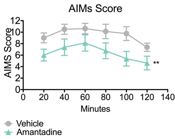
Abnormal involuntary movements (AIMS) in the 6-OHDA Model. Once daily administration of amantadine (40 mg/kg) significantly reduces AIMs score on day 12 of dosing. Data are mean ± SEM; **p<0.01 compared to vehicle. Scores represent the sum of axial, limb and orolingual AIMs at multiple time points over a two hour observation period (N=8-9).
In addition to AIMs testing, Forepaw Adjusting Steps (FAS) testing was conducted over a series of trials to quantify the number of adjusting steps for both paws in the forehand and backhand directions of movement. This test is used to assess motor deficits in the forelimbs. Lesioned rats show a unilateral motor deficit in adjusting steps compared to unlesioned animals. Chronic treatment with L-DOPA can improve this function, and it is important to determine if a treatment interferes with this L-DOPA effect.
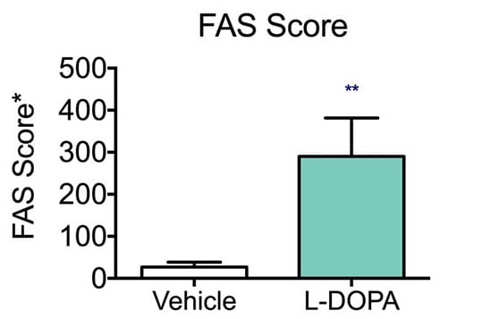
Forelimb Adjusting Step (FAS), A Measure of Parkinsonism in the 6-OHDA Model. Once daily administration of L-DOPA (12 mg/kg) alleviates akinesia as demonstrated by improved performance in the Forepaw Adjusting Step (FAS) test. Data are mean ± SEM; **p<0.01 compared to vehicle (N=8-9).
The 6-OHDA Hemiparkinsonian Lesion model is only conducted in rats. The studies are normally about 2 months in duration. Ideal group sizes are >8 animals. In addition to FAS and AIMs, histological endpoints such as the evaluation of tyrosine hydroxylase (a marker of dopaminergic neurons) positive immunohistochemistry in the striatum and substantia nigra or cell counts of TH-positive cells in the substantia nigra can be conducted.


