Allergic Contact Hypersensitivity - Atopic Dermatitis Mouse Model
Discover how Melior’s unique phenotypic screening platforms can uncover the untapped value of your candidate therapeutic
Contact hypersensitivity models mimic contact dermatitis aspects of immune function. Many cutaneous disorders are characterized by inflammation involving different immune signalling pathways. Accordingly there are variances of contact hypersensitivity animal models of atopic dermatitis that use different haptens to activate different immune pathways.
The immune reaction induced by the method described below is regarded as an atopic dermatitis mouse model. It is associated with epidermal swelling at the site of challenge. Melior has established models of allergic contact dermatitis hypersensitivity using oxazolone, DNFB and other haptens which vary in the degree to which they involve Th1 versus Th2 pathways. The localized inflammatory responses can be attenuated by corticosteroids and topical agents used to treat atopic dermatitis.
Allergic Contact Hypersensitivity - Atopic Dermatitis Mouse Model
Contact us today for more information about our bespoke research models and to discuss how we can help you answer your unique research questions.


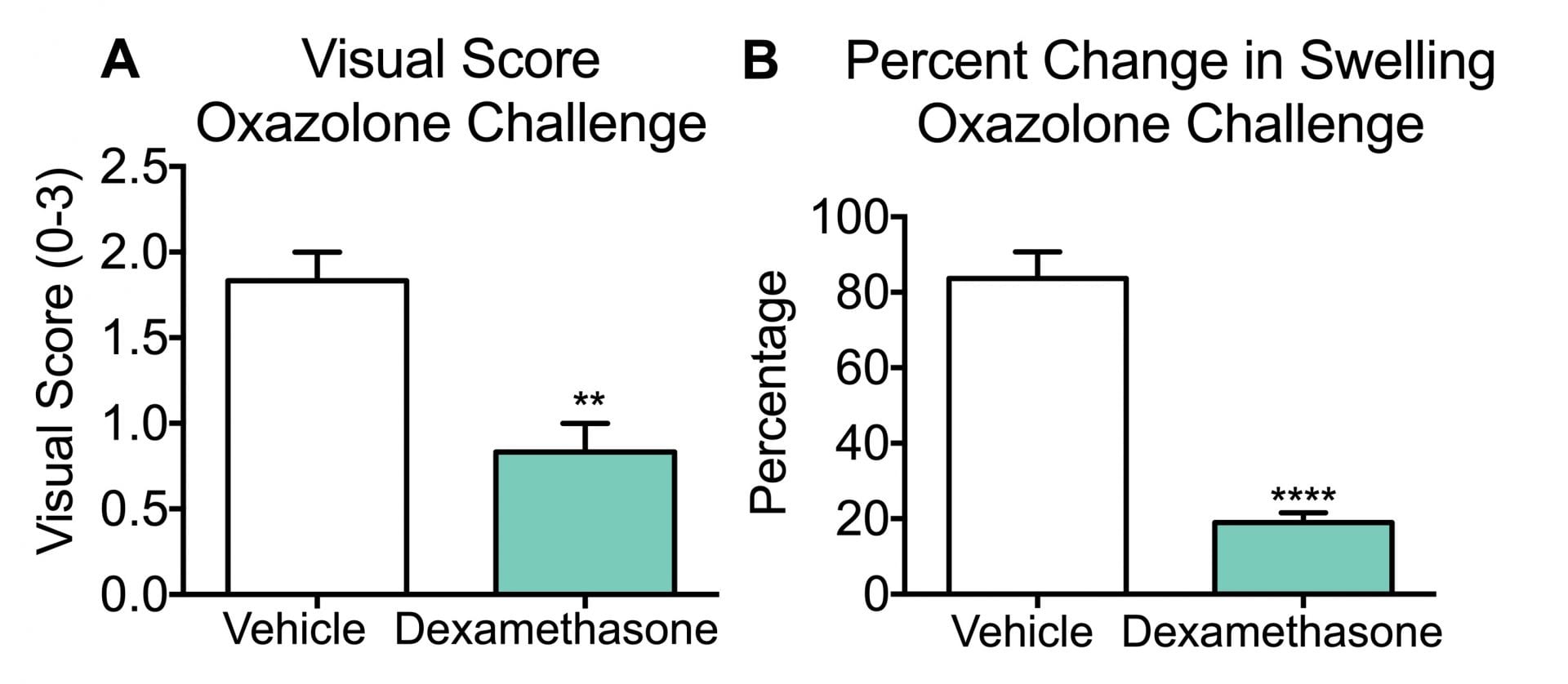
Five days after baseline and initial sensitization to either oxazolone (A and B) or DNFB (C and D), CD-1 male mice were administered the compound and challenged with hapten. Any resulting sensitivity (i.e: swelling and/or redness) is mechanically measured and visually scored 24 hours after the challenge.
The figures illustrate the visual scoring (0 = no swelling or redness; 1 = minor swelling or redness; 2 = moderate swelling and some redness; 3 = severe swelling and prominent redness) and the tissue thickness after the challenge. Both agents produced an allergic reaction measured as tissue swelling and visual score. Dexamethasone significantly attenuated the allergic reaction compared to vehicle-treated animals (***p<0.001). Data are mean ± SEM; ***p<0.001 compared to vehicle (N=8).
The figures illustrate the visual scoring (0 = no swelling or redness; 1 = minor swelling or redness; 2 = moderate swelling and some redness; 3 = severe swelling and prominent redness) and the tissue thickness after the challenge. Both agents produced an allergic reaction measured as tissue swelling and visual score. Dexamethasone significantly attenuated the allergic reaction compared to vehicle-treated animals (***p<0.001). Data are mean ± SEM; ***p<0.001 compared to vehicle (N=8).
As described above this is typically run as a 1 week model and typically involves groups sizes on the order of 8 to 10 mice depending upon effect size expected for a test article. Most commonly test articles are administered acutely (one time).
