Subthalamic Nucleus (STN) Recording In Vivo
Discover how Melior’s unique phenotypic screening platforms can uncover the untapped value of your candidate therapeutic
In Parkinson’s disease patients, it has been demonstrated that neurons in the subthalamic nucleus (STN) show an atypical bursting pattern. Reduction of STN bursting by lesions or deep-brain stimulation has been demonstrated to ameliorate Parkinsonian symptoms in humans and monkeys (see e.g. Patel et al, 2003; Brain 126:1136-45).
STN burst firing has also been demonstrated in animal models in which nigro-striatal dopaminergic pathways have been chemically ablated, such as the rat 6-OHDA-lesion model. This model may thus be valuable for evaluating therapeutic treatments for Parkinson’s disease.

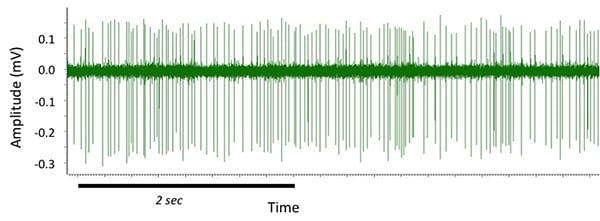
Acute recording from an STN neuron using stereotaxically implanted metal microelectrode in an anesthetized normal rat. Note relatively steady firing.

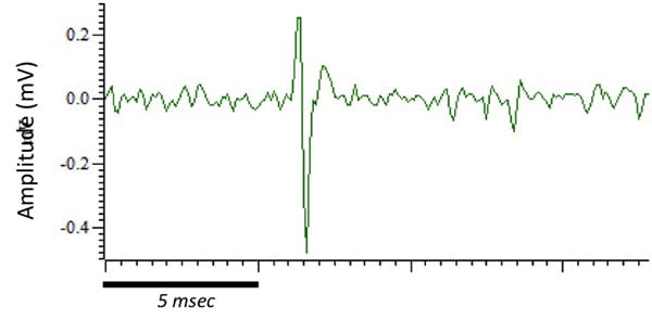
Isolated spike from an STN neuron in a 6-OHDA-lesioned rat. Spike has characteristic biphasic shape with slight “tail”.
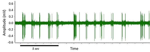
Clear bursting activity was observed in STN cells in this rat that received a dopaminergic lesion produced by stereotaxic injection of 6-OHDA into the medial-forebrain bundle (MFB). The lesion was confirmed by apomorphine-induced rotations 4 weeks later. Bursting parameters are quantified off-line.
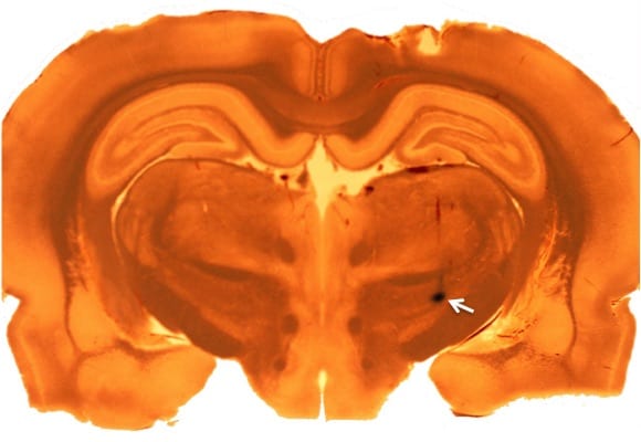
At the end of recording, pontamine sky-blue dye was iontophoretically injected into the recording site (white arrow) to confirm recording from the STN.
References
Patel NK, Heywood P, O’Sullivan K, McCarter R, Love S, Gill SS. (2003). Unilateral subthalamatomy in the treatment of Parkinson’s disease. Brain 126:1136-45.


