
The K7M2 Osteosarcoma Tumor Model
Osteosarcoma Model: Key Insights, Treatment Options, and the Role of Mouse Models in Research
Osteosarcoma (OS) is a rare and aggressive bone cancer that predominantly affects children and young adults. This malignancy is commonly found in the long bones, particularly around the knee, and is known for its rapid metastasis, primarily to the lungs. Despite advancements in surgery and chemotherapy, prognosis for patients with metastatic or recurrent osteosarcoma remains poor. This highlights the urgent need for novel treatment options, and the development of an osteosarcoma model plays a critical role in advancing research and therapeutic strategies.
Treatment Options for Osteosarcoma
The standard treatment approach for osteosarcoma typically involves neoadjuvant chemotherapy, followed by surgery, and adjuvant chemotherapy. Chemotherapeutic regimens often include drugs like methotrexate, doxorubicin, and cisplatin. While these treatments have improved survival rates, side effects such as cardiotoxicity, nephrotoxicity, and secondary cancers remain significant concerns. For patients with metastatic osteosarcoma or those who experience relapse, the prognosis is still grim, with survival rates often falling below 30%. New treatment modalities, including immunotherapy, targeted therapy, and gene editing, are being investigated, but their effectiveness in clinical settings remains under study.
Unmet Medical Needs in Osteosarcoma Treatment
Despite current therapies, there are major unmet medical needs in osteosarcoma care. Metastatic and recurrent osteosarcoma still has limited effective treatments, and the development of resistance to chemotherapy is a major challenge. Additionally, long-term side effects from chemotherapy significantly impact patients’ quality of life. There is a critical need for more effective, targeted therapies with fewer long-term consequences, particularly as osteosarcoma remains a rare disease, limiting clinical trials and research focus.
The Role of the Mouse Model of Osteosarcoma
Animal models are indispensable for understanding the biology of osteosarcoma and for testing new therapies. The K7M2 syngeneic mouse model of osteosarcoma is a widely used tool in preclinical research. This model, where murine osteosarcoma cells are implanted into immunocompetent Balb/C mice, closely replicates key aspects of human osteosarcoma, including tumor progression, metastasis, and chemotherapy resistance. The K7M2 osteosarcoma model has been instrumental in evaluating innovative treatment options like immunotherapy and targeted drugs, helping to refine therapeutic strategies before clinical trials. Melior runs the K7M2 osteosarcoma model as either a subcutaneous tumor model or an orthotopic model in which cells are engrafted into the tibia.
Tailor-made Services: Designed with You in Mind
Initiate your K7M2 tumor model study quickly with a bespoke design to suit your needs. In as little as 3-4 weeks, we help you go from design to results with our flexible, custom services.
Our bespoke analyses include:
- Tissue collection for immune cell analysis and profiling
- Whole blood, spleen, and lymph node analysis
- General observations, including pain analysis
- Histology and IHC
- Luciferase assay
- IVIS imaging
- FACS analysis
- PK studies
Ready to get started or looking for a custom model?
We offer face-to-face Zoom calls, ad-hoc consultations, and customized study proposals. Take advantage of a direct line to experienced project managers and our hands-on approach to revision support.
Expand Your Applications with immuno-theraTRACEⓇ
With our oncology platform, immuno-theraTRACE®, you can test your immunotherapeutic across 9 syngeneic models. This helps you determine the best target cancer type to advance your immunotherapy.
This model is included in our immuno-theraTRACE® oncology platform


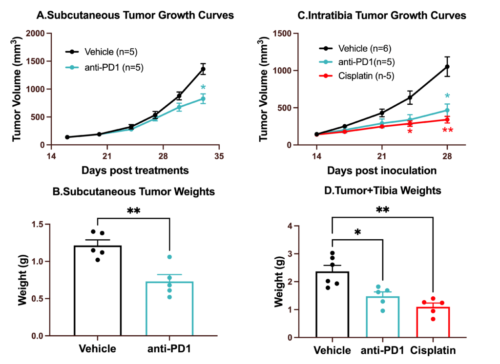
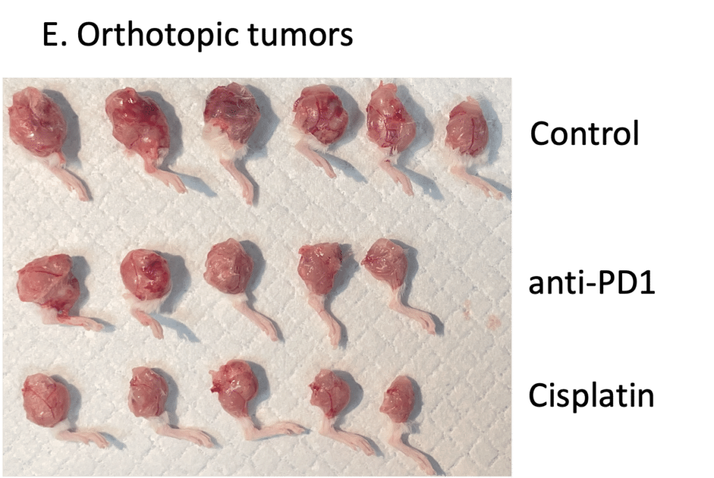
Chemotherapy and immune check point inhibitor validations in mouse K7M2 subcutaneous and orthotopic syngeneic mouse model. 1x 10⁶ K7M2 cells were subcutaneously or orthotopically (intratibial) injected into Balb/C mice. Once the tumor size reached ~150mm3, mice were randomized into groups of vehicle (normal saline), anti PD-1 (12.5mg/kg, IP weekly) and cisplatin (4 mg/kg, IP weekly). Subcutaneous tumor growth curve and tumor weights are shown in A and B. Orthotopic tumor growth curves, tumor+tibia weights and tumors are shown in C, D and E. Data are mean ± SEM. . Data are mean ± SEM.
Publications
Citations
- Jafari, F., Javdansirat, S., Sanaie, S., Naseri, A., Shamekh, A., Rostamzadeh, D., & Dolati, S. (2020). Osteosarcoma: A comprehensive review of management and treatment strategies. Annals of diagnostic pathology, 49, 151654, 13(1), 927.
- Ottaviani, G., & Jaffe, N. (2010). The epidemiology of osteosarcoma. Pediatric and adolescent osteosarcoma, 3-13.BMC cancer, 21, 1-12.
- Walia, M. K., Castillo‐Tandazo, W., Mutsaers, A. J., Martin, T. J., & Walkley, C. R. (2018). Murine models of osteosarcoma: a piece of the translational puzzle. Journal of cellular biochemistry, 119(6), 4241-4250.BioMed Research International, 2021(1), 6626851.
- Kuo Jiang, Qianfeng Zhang, Yong Fan, Jia Li, Jitao Zhang, Wentao Wang, Jinzhu Fan, Yunshan Guo, Shichang Liu, Dingjun Hao, Yongxiang Wang, Lei Wang & Lequn Shan. Cell Death Discovery (2022)8:117 ; https://doi.org/10.1038/s41420-022-00923-8, 36(11), 931-940.